|
The Achilles tendon is the largest tendon in the body. It is
formed by a confluence of the tendons of the gastrocnemius and
soleus muscles. It inserts onto the posterior aspect of the
calcaneum. It assists in plantar flexion of the foot. The
Achilles tendon does not have a synovial sheath. On MRI
-
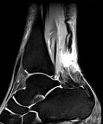
Partial Rupture:
-
There is partial continuity of a
portion of the tendon fibres on at least one sagittal
section. There is no tendinous gap.
-
The tendon may be thickened and usually
exhibits focal areas of intermediate signal intensity on
the T1W images and increased signal intensity on the T2W
images due to edema and/or hemorrhage.
-
It may be difficult to differentiate
between tendinitis and partial tears as the two often
coexist. In uncomplicated chronic tendinitis there is
focal or diffuse thickening of the tendon without
increased intrasubstance signal intensity.
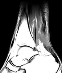
- Complete Rupture:
-
There is discontinuity of the tendon
(unless the tendon edges are overlapping) with
intervening fluid, fat or hemorrhage.
-
The proximal fragment is retracted with
fraying. The distal fragment is lax and buckled.
Sagittal images help in ascertaining the distance
between the two fragments.
-
Hemorrhage, edema and inflammation may
be seen in the peritendinous soft tissues. There may be
fluid collection in the paratenon anterior to the
tendon.
|