|
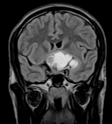
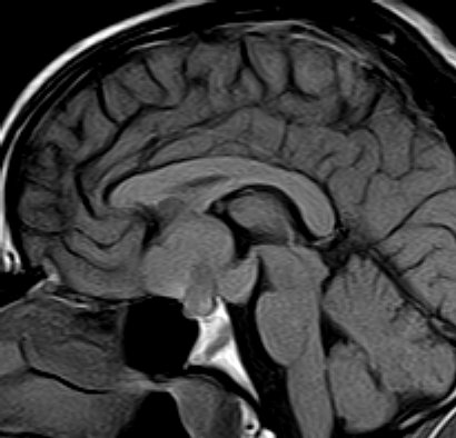
There is
evidence of a lobulated extra-axial mass lesion in the
suprasellar cistern. The lesion is predominantly hyperintense on
the T1W images and heterogeneously hyperintense on the T2W and
FLAIR images. The optic chiasm, proximal optic nerves and the
pituitary stalk cannot be identified separately from this
lesion. However, the pituitary gland is well identified.
Craniopharyngiomas are epithelially derived neoplasms that
usually occur in the suprasellar cistern. Occasionally they
occur in the sella or in the third ventricle. They constitute
approximately 3% of all intracranial tumors and show no sex
predilection. Craniopharyngiomas are hormonally inactive
lesions. 50% of these lesions occur in childhood or adolescence,
with a peak incidence between 5 and 10 years of age. A second
smaller peak is seen in the sixth decade. Patients may present
with headaches and/or visual disturbances. The lesions usually
exhibit a heterogeneous appearance with presence of a cystic
and/or solid component. It may be hyperintense on both T1W and
T2W images. The lesions may encase nearby cerebral vasculature.
The solid portion may be calcified. On contrast the solid
portion usually enhances.
|