|
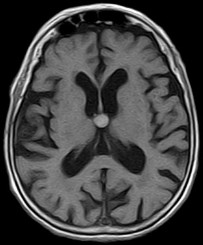
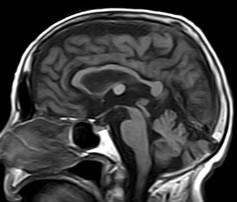
Colloid cysts are benign, epithelial lined lesions,
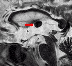
usually located antero-superior to the third ventricle
(between the columns of the fornices). They supposedly
originate due to an infolding of the neuroepithelium. These
cysts may also occur in the choroid plexus of the lateral
ventricles, subarachnoid space and brain parenchyma. They
comprise approximately 2% of all glial neoplasms. Patients may
present with headaches, sudden transient paralysis of both
lower extremities, urinary incontinence, personality changes
and/or dementia.
-
On MRI
-
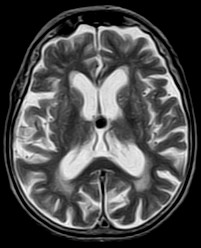
These lesions may be either hypointense or
hyperintense on either T1W or T2W images. The variability is
due to it's contents. They may contain mucoid material,
blood/hemosiderin, macrophages, cholesterol crystals, CSF
and various ions (Na, Mg, Ca, Cu, Si, Al, Fe, P).
-
Occasionally they may contain serous fluid
and follow CSF signal characteristics.
-
The lesions are thin walled and peripheral
enhancement is common.
|